
Treatment for staphylococcal pneumonia depends on if this process is categorized into either MRSA or MSSA. A procalcitonin level can also be drawn at the time of treatment in order to guide antibiotic efficacy and aid in antibiotic stewardship. An MRSA swab may also be utilized to assess the patient's colonization status for MRSA and, therefore, further the suspicion of this process. However, if the patient can produce an adequate sample, 82% were found to be diagnostic and could be used to guide antibiotic therapy. A sputum sample can be obtained, but a 2005 study noted that only 17% of the study population were able to produce an adequate sample. If the clinical syndrome supports the diagnosis of pneumonia, but the chest radiograph does not reveal pathology, a computed tomography (CT) scan can be performed to further evaluate for pathology in the lungs.įindings on CT scan may show a developing lobar infiltrate or cavitary lesion that chest radiograph may not reveal. The infiltrate on a chest radiograph can show lobar infiltrate or in severe patients can show cavitary lesions and empyema. The gold standard for diagnosing pneumonia with the appropriate clinical suspicion is the presence of infiltrate on a chest radiograph. A complete blood count (CBC) will likely show leukocytosis with a neutrophilic predominance.

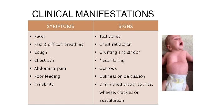
Initial evaluation for staphylococcal pneumonia starts with the same foundational workup for those suspected of having any etiology of CAP. Unfortunately, there is no specific constellation of history and physical exam findings that can effectively guarantee with certainty a diagnosis of pneumonia, but the above findings do further the clinical suspicion of this disease process. Percussion and tactile fremitus can also be utilized if there is a concern for pleural effusion. Physical exam should include a complete multisystem evaluation, but the focus should be given to the cardiopulmonary examination to assess for signs of valvular murmur, respiratory distress, tachypnea, or inspiratory crackles in a lobar pattern. In the appropriate clinical scenario, evaluation for IV drug use should be ascertained as tricuspid valve endocarditis from IV drug use can lead to septic emboli to the lungs and cause S. Employment status and recent hospitalizations can be ascertained to raise clinical suspicion for colonization with S. Evaluation for sick contacts, recent travel, ongoing or recent infection with influenza can also help delineate those who may have a higher predisposition to developing S. However, this finding is less reliable in the elderly. Eighty percent of patients with pneumonia can be febrile. When evaluating the need for hospitalization of the patient, care should be taken to assess the onset of symptoms, duration of symptoms, as well as associative symptoms of fever, chills, dyspnea, or productive cough. Therefore, a thorough multisystem history and physical should be performed. aureus infection can preclude patients to a wide range of infectious processes.


 0 kommentar(er)
